Shoulder Anatomy Diagram / Shoulder Anatomy Shoulderbuzz. The shoulder has an incredible range of motion, but this means that it is also very prone to injury.the shoulder can easily slip out of alignment by a few millimeters, become weak due to regular wear and tear, or become completely dislocated during a fall. Sechrest, md narrates an animated tutorial on the basic anatomy of the shoulder. It is one of the most mobile joints in the human body, at the cost of joint stability. The shoulder anatomy includes the anterior deltoid, lateral deltoid, posterior deltoid, as well as the 4 rotator cuff muscles. The shoulder joint (glenohumeral joint) is a ball and socket joint between the scapula and the humerus.it is the major joint connecting the upper limb to the trunk.
Most people with rotator cuff injuries can recover with rest and physical therapy. The muscles of the neck run from the base of the skull to the upper back and work together to bend the head and. Shoulder pain, instability and, in some cases, a feeling of grinding, locking or catching while moving the shoulder. Sechrest, md narrates an animated tutorial on the basic anatomy of the shoulder. The shoulder joint (glenohumeral joint) is a ball and socket joint between the scapula and the humerus.it is the major joint connecting the upper limb to the trunk.
Schultermuskulatur Diagramm Diagramm Schultermuskulatur Precautions To Take In Handling A Hurt Dog D Muscle Anatomy Human Body Anatomy Shoulder Anatomy from i.pinimg.com The shoulder joint is not very stable, and it may be easily dislocated as the anatomy is conducive to that and the soft tissues around the joint are. The muscles of the shoulder support and produce the movements of the shoulder girdle.they attach the appendicular skeleton of the upper limb to the axial skeleton of the trunk. Two joints are at the shoulder. Rotator cuff injuries are very common, affecting over 3 million people in the united states every year. The shoulder has about eight muscles that attach to the scapula, humerus, and clavicle. Other important bones in the shoulder include: 17 photos of the diagram of shoulder muscles and tendons. Plus, exercises for training them.
The most flexible joint in the entire human body, our shoulder joint is formed by the union of the humerus, the scapula (or shoulder blade), and the clavicle (or collarbone).
Rotator cuff and shoulder anatomy. Formerly called tendinitis, this is inflammation or irritation of a tendon that attaches to a bone. The most flexible joint in the entire human body, our shoulder joint is formed by the union of the humerus, the scapula (or shoulder blade), and the clavicle (or collarbone). It is one of the most mobile joints in the human body, at the cost of joint stability. The shoulder bones include the scapula (shoulder blade), humerus (upper arm bone), and clavicle (collarbone). The joints between these bones are flexible and allow for a wide range of motion. The anterior shoulder pain usually develops when injury or inflammation occurs in the tendons that are attached to the shoulder joint. The shoulder has about eight muscles that attach to the scapula, humerus, and clavicle. Other important bones in the shoulder include: These muscles form the outer shape of the shoulder and underarm. Plus, exercises for training them. Most people with rotator cuff injuries can recover with rest and physical therapy. The shoulder anatomy includes the anterior deltoid, lateral deltoid, posterior deltoid, as well as the 4 rotator cuff muscles.
The shoulder joint is formed where the humerus (upper arm bone) fits into the scapula (shoulder blade), like a ball and socket. An example of shoulder flexion the muscles of the rotator cuff are common sites of injury in adults, particularly among people who. The joints between these bones are flexible and allow for a wide range of motion. Numerous muscles help stabilize the three joints of. These muscles form the outer shape of the shoulder and underarm.
Shoulder Muscles Anatomy High Resolution Stock Photography And Images Alamy from c8.alamy.com 🤔 the acetabulofemoral joint , commonly called the hip joint , scientifically termed is located in between the pelvis and the femur of the legs. The muscles of the shoulder support and produce the movements of the shoulder girdle.they attach the appendicular skeleton of the upper limb to the axial skeleton of the trunk. Anatomy and injuries of the shoulder anatomical chart. This diagram depicts shoulder muscle diagram. In this episode of eorthopodtv, orthopaedic surgeon randale c. Learn about these muscles, their origin and insertion points, and their functional anatomy. The shoulder joint is formed where the humerus (upper arm bone) fits into the scapula (shoulder blade), like a ball and socket. Localized anatomy of the rotator cuff and shoulder.
The bones of the shoulder are:
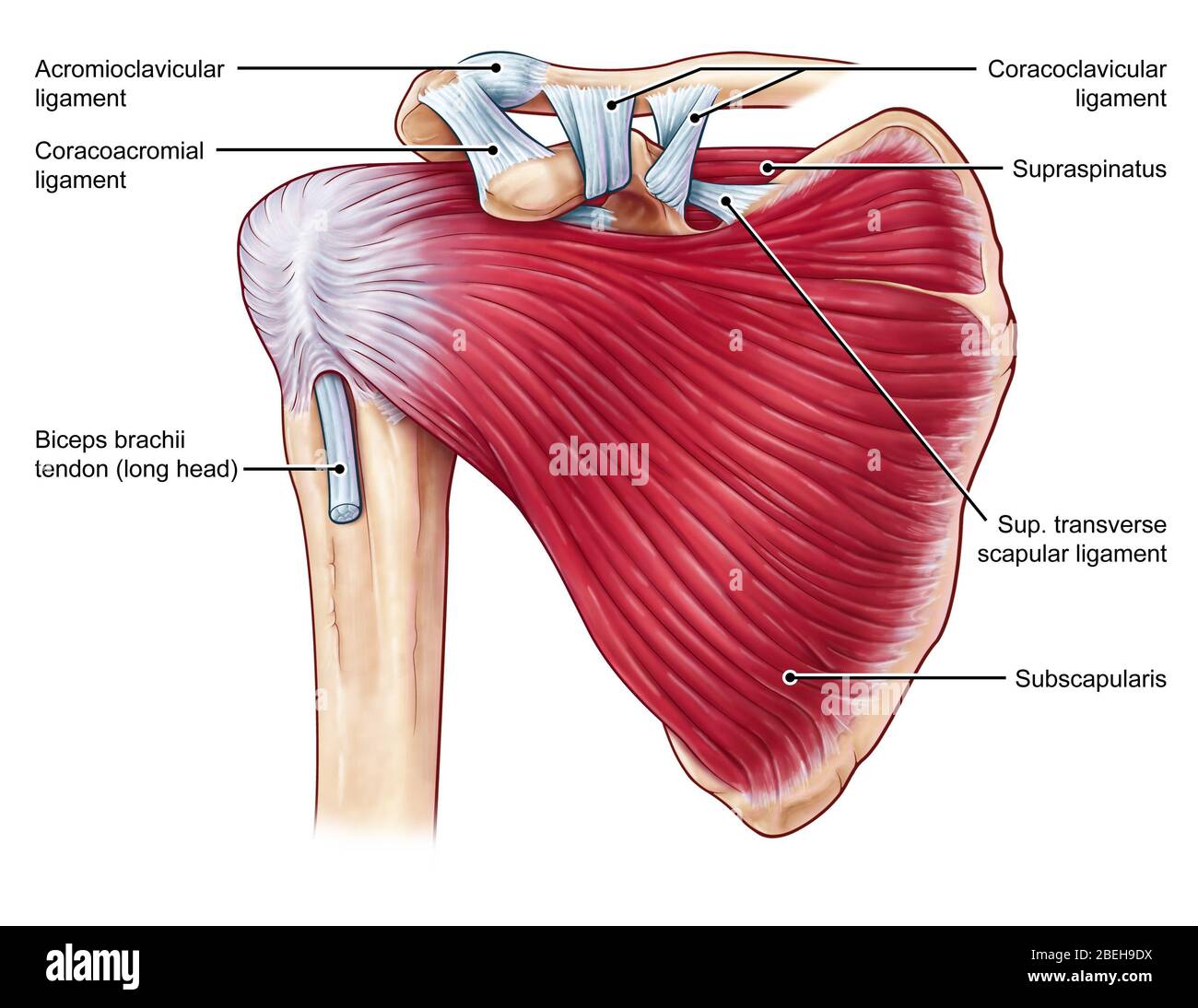
The shoulder is a complex combination of bones and joints where many muscles act to provide the widest range of motion of any part of the body. Shoulder pain anatomy map / anatomy of neck and shoulders anatomy drawing diagram. The shoulder is made up of 3 bones: The following is an overview of the shoulder muscle anatomy. A second joint in the shoulder is the junction of the collar bone with the shoulder blade, called the. The different types of connective tissues in the shoulder are bone, articular cartilage, ligaments, joint capsules, and bursa (see gross anatomy). The shoulder joint is formed where the humerus upper arm bone fits into the scapula shoulder blade like a ball and socket. In this episode of eorthopodtv, orthopaedic surgeon randale c. However, more serious injuries, such as complete rotator cuff tears, may require surgical repair. Test your knowledge of the clavicle, scapula and humerus with our labeled diagram exercises and quizzes! Plus, exercises for training them. For that reason, and because of the dexterity of the shoulder joint itself, the musculature of the shoulder is complex, ranging from massive prime mover muscles to finer stabilizer and fixator muscles. Illustration of the shoulder anatomy and labrum.
Plus, exercises for training them. Two joints are at the shoulder. Shoulder pain anatomy map / anatomy of neck and shoulders anatomy drawing diagram. Male shoulder ligaments and biceps muscles isolated in skeleton labeled chart on white labeled human anatomy diagram of male shoulder ligaments, connective tissue and biceps muscles isolated within the skeletal system frontal anterior view on a white background. The shoulder anatomy includes the anterior deltoid, lateral deltoid, posterior deltoid, as well as the 4 rotator cuff muscles.
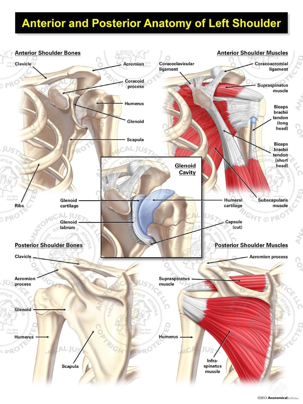
Anterior And Posterior Anatomy Of The Left Shoulder Anatomical Justice from anatomicaljustice.com Shoulder muscle anatomy neck muscle anatomy shoulder muscles supraspinatus muscle muscle fascia muscle diagram human body organs anatomy images latissimus dorsi. The muscles of the neck run from the base of the skull to the upper back and work together to bend the head and. The humerus is the bone of the arm that articulates with the scapula proximally and with the radius and the ulna distally. Localized anatomy of the rotator cuff and shoulder. The muscles of the shoulder bridge the transitions from the torso into the head/neck area and into the upper extremities of the arms and hands. Formerly called tendinitis, this is inflammation or irritation of a tendon that attaches to a bone. The shoulder joint is formed where the humerus upper arm bone fits into the scapula shoulder blade like a ball and socket. Deltoides triangular refers to the front head of the.
The anterior shoulder pain usually develops when injury or inflammation occurs in the tendons that are attached to the shoulder joint.
Bones in shoulder, ligaments of the shoulder joint, parts of the shoulder joint, shoulder anatomy, shoulder joints and muscles, shoulder structure anatomy, shoulder tendon anatomy, shoulder tendons ligaments, human muscles, bones in shoulder, ligaments of the shoulder joint, parts of. The muscles of the neck run from the base of the skull to the upper back and work together to bend the head and. 🤔 the acetabulofemoral joint , commonly called the hip joint , scientifically termed is located in between the pelvis and the femur of the legs. 17 photos of the diagram of shoulder muscles and tendons. It is one of the most mobile joints in the human body, at the cost of joint stability. Ac joint is a diathrodial joint with a fibrocartilaginous disk. The different types of connective tissues in the shoulder are bone, articular cartilage, ligaments, joint capsules, and bursa (see gross anatomy). Shoulder pain anatomy map / anatomy of neck and shoulders anatomy drawing diagram. Human anatomy diagrams show internal organs, cells, systems, conditions, symptoms and sickness information and/or tips for healthy living. Illustration of the shoulder anatomy and labrum. The most flexible joint in the entire human body, our shoulder joint is formed by the union of the humerus, the scapula (or shoulder blade), and the clavicle (or collarbone). The joints between these bones are flexible and allow for a wide range of motion. Shoulder pain, instability and, in some cases, a feeling of grinding, locking or catching while moving the shoulder.
Share this post
0 Response to "Shoulder Anatomy Diagram / Shoulder Anatomy Shoulderbuzz"

0 Response to "Shoulder Anatomy Diagram / Shoulder Anatomy Shoulderbuzz"
Posting Komentar